Mississippi Science BIO.1C
Standard Cluster
Students will relate the diversity of organelles to a variety of specialized cellular functions. Develop and use models to explore how specialized structures within cells (e.g., nucleus, cytoskeleton, endoplasmic reticulum, ribosomes, Golgi apparatus, lysosomes, mitochondria, chloroplast, centrosomes, and vacuoles) interact to carry out the functions necessary for organism survival. Investigate to compare and contrast prokaryotic cells and eukaryotic cells, and plant, animal, and fungal cells. Contrast the structure of viruses with that of cells and explain why viruses must use living cells to reproduce.
2026 Mississippi College- and Career-Readiness Standards for Science · Cells as a System
Cluster contents
Standards in This Cluster
BIO.1C is a cluster heading. These are the individual standards under it.
- BIO.1C.1
Develop and use models to explore how specialized structures within cells (e.g., nucleus, cytoskeleton, endoplasmic reticulum, ribosomes, Golgi apparatus, lysos...
- BIO.1C.2
Investigate to compare and contrast prokaryotic cells and eukaryotic cells, and plant, animal, and fungal cells.
- BIO.1C.3
Contrast the structure of viruses with that of cells and explain why viruses must use living cells to reproduce.
Teacher's field guide
What This Cluster Means
What Students Need to Do
- Students make models that show how cell parts work together. They compare bacteria with plant, animal, and fungal cells. They explain why viruses need living cells to make more viruses.
What Mastery Looks Like
- A student can trace a protein from a ribosome through the ER and Golgi, with arrows that show what happens at each step. They can sort cells using nuclei, cell walls, and chloroplasts. They can explain that a virus has no ribosomes of its own and must use a host cell to make its parts.
Common Misconceptions
- Students may think every cell has a nucleus, or that fungi have chloroplasts because they have cell walls. They may draw organelles as separate parts with no materials moving between them. Some think viruses are cells that divide on their own.
How to Assess It
- Give students a five-row exit ticket labeled bacterium, plant, animal, fungus, and virus. Have them mark which have ribosomes, nuclei, cell walls, and chloroplasts, then draw the ribosome-to-ER-to-Golgi route in a plant cell.
Lesson moves
Ways to Teach It
Give pairs paper ribosome, ER, Golgi, and membrane cards; have them pass a labeled protein token along the route and explain each handoff.
Ask students to write why a flu virus needs a host cell to make copies, while a bacterium can reproduce when it has nutrients.
Hand teams picture cards of bacteria, leaf cells, cheek cells, yeast, and viruses; have them sort by nucleus, cell wall, and chloroplast.
Show leaf, bread yeast, and cheek cell micrographs; ask which cells can make sugar from light and which need another source.
Free download
Printable BIO.1C Worksheet
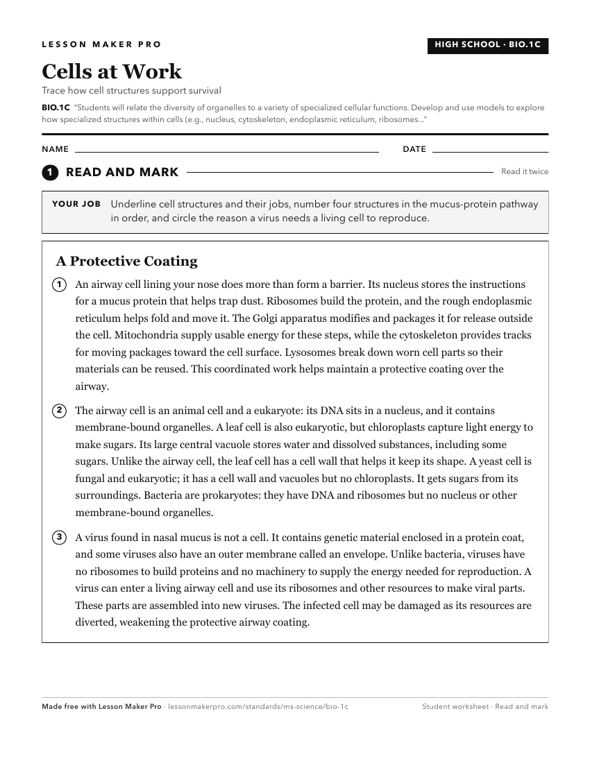
A ready-to-print activity worksheet aligned to BIO.1C, with an answer key for the teacher on its own page. No account needed.
PDF, US Letter, prints cleanly in black and white.
Download the worksheetKeep exploring
Related Standards
- FB.3
Students will demonstrate an understanding of how the structure of living organisms supports the essential functions of life.
- L.4.1
Students will demonstrate an understanding of the organization, functions, and interconnections of the major human body systems.
- HAP.2
Students will demonstrate an understanding of the relationship of cells and tissues that form complex structures of the body.
- BIO.1A
Students will demonstrate an understanding of the characteristics of life and biological organization.
Turn this cluster into a lesson
Grade, subject, topic, and the complete cluster are prefilled. Create one free, no account needed.
Also for this cluster:Make a WorksheetMake a QuizMake a Sub Lesson