Indiana Science HS-AP6-3
The Standard
Make and/or use a model of the anatomy of the ear. Construct an explanation for sensorineural and conductive hearing loss using the basic structure and function of the ear.
Indiana Academic Standards for Science (2023) · Integration and Coordination, Somatic and Special Senses
Teacher's field guide
What This Standard Means
What Students Need to Do
- Students use a model to trace sound through the ear canal, eardrum, small bones, cochlea, and auditory nerve. They explain how a blockage differs from damage to the cochlea or nerve.
What Mastery Looks Like
- A student labels the main ear parts and shows where vibrations become nerve signals. Given earwax buildup or damaged cochlear hair cells, they name the type of hearing loss and explain which step fails.
Common Misconceptions
- Students may think all hearing loss comes from a damaged auditory nerve. They may also think the cochlea just passes vibrations along, rather than turning them into nerve signals.
How to Assess It
- On an unlabeled ear diagram, have students trace the path of sound and explain whether earwax buildup and damaged cochlear hair cells cause conductive or sensorineural hearing loss.
Lesson moves
Ways to Teach It
Build a cardboard ear path with a paper canal, foil eardrum, craft-stick ossicles, and clay cochlea; block the canal with cotton.
Ask students to write why clearing earwax might restore hearing but would not repair damaged cochlear hair cells.
Give students cards showing earwax, a torn eardrum, damaged hair cells, and an injured auditory nerve; have them sort and justify each case.
Show hearing aid and cochlear implant diagrams; ask how amplified sound differs from signals sent directly to the auditory nerve.
Free download
Printable HS-AP6-3 Worksheet
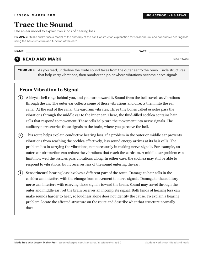
A ready-to-print activity worksheet aligned to HS-AP6-3, with an answer key for the teacher on its own page. No account needed.
PDF, US Letter, prints cleanly in black and white.
Download the worksheetKeep exploring
Related Standards
- HS-AP3-1
Develop a model to illustrate the microscopic structure, development of, maintenance of, and function of compact and spongy bone.
- HS-AP6-2
Make and/or use a model of the anatomy of the eye; then construct an explanation for hyperopia, myopia and astigmatism using the model.
- HS-AP5-1
Develop a model that illustrates the structural components and functional subdivisions of the nervous system.
- HS-AP1-2
Develop and use a model to illustrate the hierarchical organization of structural body systems that provide specific functions within the Human Body.
Turn this exact standard into a lesson
Grade, subject, topic, and the complete standard are prefilled. Create one free, no account needed.
Also for this standard:Make a WorksheetMake a QuizMake a Sub Lesson