Indiana Science HS-AP5-3
The Standard
Compare and contrast the actions, origins, and pathways of nerve fibers in the parasympathetic and sympathetic divisions of the autonomic nervous system.
Indiana Academic Standards for Science (2023) · Integration and Coordination, the Nervous System
Teacher's field guide
What This Standard Means
What Students Need to Do
- Students compare where sympathetic and parasympathetic signals start and the routes they take to organs. They predict how each changes heart rate, pupil size, and digestion.
What Mastery Looks Like
- Students locate the sympathetic origin in the thoracic and lumbar spinal cord, and the parasympathetic origins in the brainstem and sacral spinal cord. They draw short first fibers to ganglia near the spine for sympathetic signals, and long first fibers to ganglia near organs for parasympathetic signals. They predict which division raises heart rate and which increases digestion.
Common Misconceptions
- Students may think both divisions start in the same place or that one nerve runs straight from the spinal cord to an organ. They may think sympathetic activity turns every organ on and parasympathetic activity turns every organ off. Digestion does not follow that pattern.
How to Assess It
- Draw both nerve routes to the heart. Label each starting point, ganglion, and first fiber length, then circle the route that raises heart rate.
Lesson moves
Ways to Teach It
Use yarn on a body outline to build both heart pathways, placing paper ganglia near the spine or near the heart.
Write two sentences comparing heart rate and digestion before a speech and during a relaxed meal, naming the more active division in each.
Give pairs cards labeled brainstem, thoracic cord, sacral cord, near-spine ganglion, near-organ ganglion, short fiber, and long fiber to sequence both pathways.
Show heart-rate readings from a runner during a sprint and after rest; students name the likely more active division at each point.
Free download
Printable HS-AP5-3 Worksheet
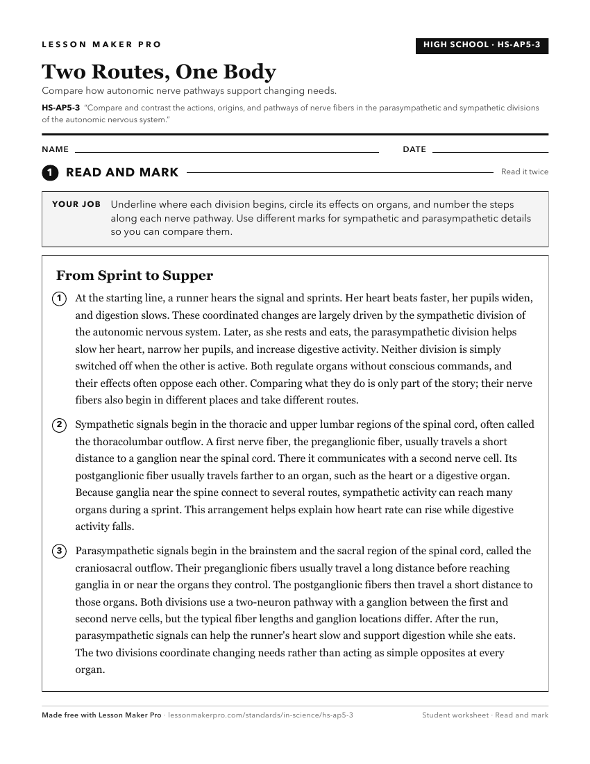
A ready-to-print activity worksheet aligned to HS-AP5-3, with an answer key for the teacher on its own page. No account needed.
PDF, US Letter, prints cleanly in black and white.
Download the worksheetKeep exploring
Related Standards
- HS-AP6-1
Compare and contrast the somatic, visceral, and special senses, the prominent sensory receptor types of each, and their functional operation.
- HS-AP5-2
Observe and identify the structure and function of the various neurons and neuroglia. Explain how the varying structures determine the specified function.
- HS-AP1-3
Compare and contrast the relationships among the various tissue types as well as the molecular and cellular composition of these tissues.
- HS-AP4-1
Compare and contrast between the structural and functional characteristics of skeletal, cardiac, and smooth muscle.
Turn this exact standard into a lesson
Grade, subject, topic, and the complete standard are prefilled. Create one free, no account needed.
Also for this standard:Make a WorksheetMake a QuizMake a Sub Lesson