Alabama Science HAP.1
The Standard
Obtain, evaluate, and communicate information to explain how differences in cellular structure (mitochondria, cytoskeletal structure, endoplasmic reticulum, cell membrane) lead to differences in the function and organization of the four tissue types (epithelial, connective, muscular, and nervous).
2023 Alabama Course of Study: Science · From Molecules to Organisms: Structures and Processes
Teacher's field guide
What This Standard Means
What Students Need to Do
- Students compare diagrams and short notes about epithelial, connective, muscle, and nervous tissues. They explain how cell membranes, mitochondria, the cytoskeleton, and ER help each tissue work and take its shape. They check whether the diagrams and notes support their claims.
What Mastery Looks Like
- Students connect membrane junctions to an epithelial barrier and fibroblast rough ER to collagen in connective tissue. They link muscle cell mitochondria and contractile fibers to movement. They explain how a neuron's membrane carries signals and its cytoskeleton supports its long axon, using diagram details as evidence.
Common Misconceptions
- Students may think only muscle cells have mitochondria, or that each tissue has organelles found nowhere else. They may confuse the cytoskeleton inside a cell with the collagen matrix outside connective tissue cells.
How to Assess It
- Hand out a labeled four-panel diagram of epithelial cells, fibroblasts, muscle fibers, and neurons. Ask students to pick two tissues and explain, using one labeled detail for each, how a cell structure supports the tissue's job.
Lesson moves
Ways to Teach It
Use clay, string, and beads to model epithelial sheets, connective tissue, muscle fibers, and neurons, then label membranes, ER, mitochondria, and cytoskeleton.
Project a neuron diagram with membrane channels and ask, 'Why would blocking these channels change nerve signaling? Cite a diagram label.'
Play tissue match: teams pair cards showing a cell feature, tissue type, and job, then defend each match with a diagram detail.
Pair a scraped-knee photo with a fibroblast diagram; have students explain how epithelial barriers and fibroblast rough ER help repair the wound.
Free download
Printable HAP.1 Worksheet
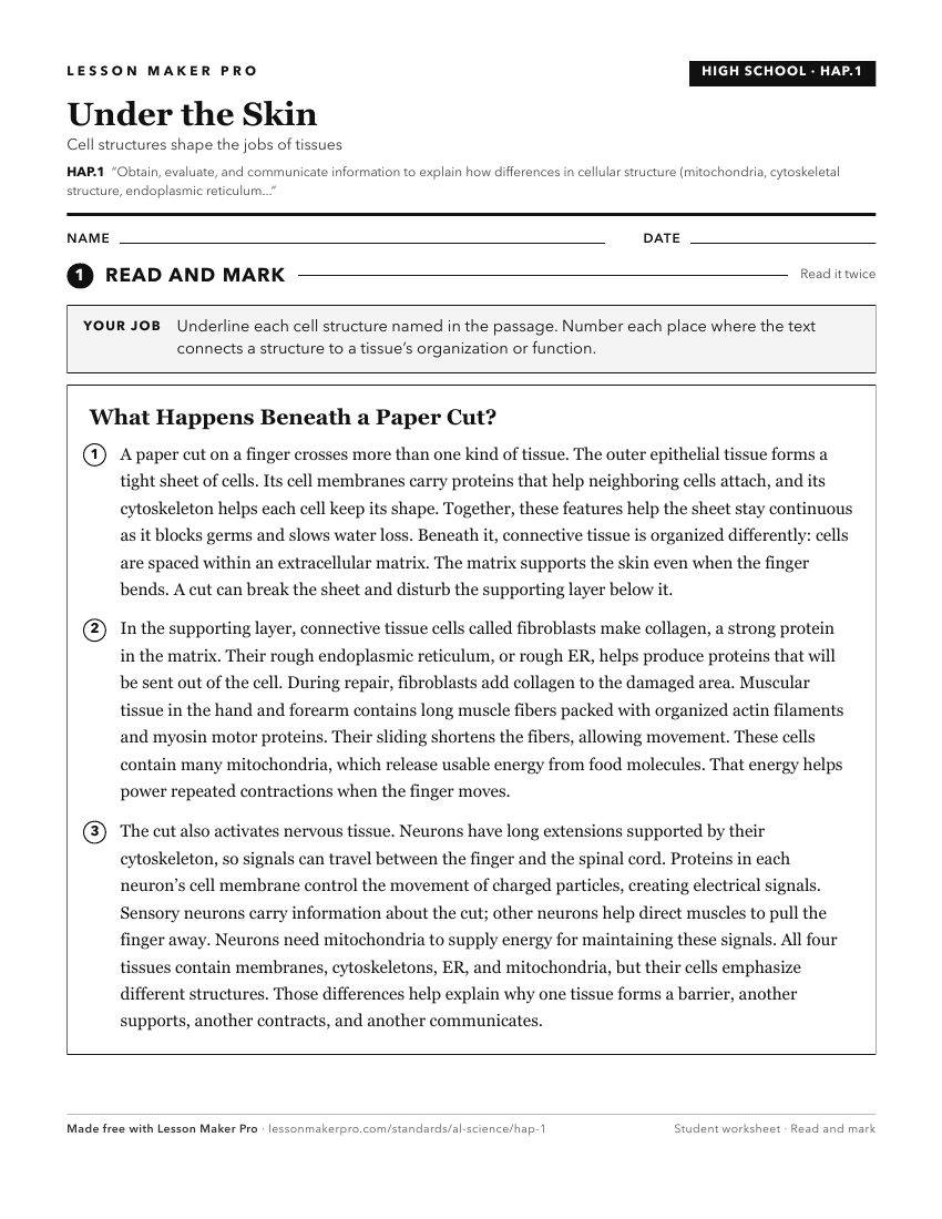
A ready-to-print activity worksheet aligned to HAP.1, with an answer key for the teacher on its own page. No account needed.
PDF, US Letter, prints cleanly in black and white.
Download the worksheetKeep exploring
Related Standards
- 7.4
Obtain, evaluate, and communicate information explaining how cells, tissues, and organs of various systems of the human body work together for specific function...
- BIO.1
Engage in evidence-based argument to relate a cell’s function to the structure, function, and diversity of its components.
- HAP.4.b
Obtain, evaluate, and communicate information to explain muscle fatigue and tone in terms of muscle cell physiology.
- HAP.8
Obtain, evaluate, and communicate information explaining how the structures of the cardiovascular system are related to its functions.
Turn this exact standard into a lesson
Grade, subject, topic, and the complete standard are prefilled. Create one free, no account needed.
Also for this standard:Make a WorksheetMake a QuizMake a Sub Lesson