Alabama Science 7.1
Standard Cluster
Develop and use a model to explain the functions of specific cell structures necessary for maintaining a stable environment, including the cell membrane, cell wall, chloroplasts, endoplasmic reticulum, golgi apparatus, mitochondria, nucleus, ribosomes, and vacuoles. a. Engage in argument from evidence to support claims of cell theory. b. Construct an explanation of how prokaryotic and eukaryotic cells differ in structure and function. c. Plan and carry out an investigation to identify and explain features of a cell’s semi-permeable membrane which enable it to control what enters and exits the cell.
2023 Alabama Course of Study: Science · From Molecules to Organisms: Structures and Processes
Cluster contents
Standards in This Cluster
7.1 is a cluster heading. These are the individual standards under it.
- 7.1.a
Engage in argument from evidence to support claims of cell theory.
- 7.1.b
Construct an explanation of how prokaryotic and eukaryotic cells differ in structure and function.
- 7.1.c
Plan and carry out an investigation to identify and explain features of a cell’s semi-permeable membrane which enable it to control what enters and exits the ce...
Teacher's field guide
What This Cluster Means
What Students Need to Do
- Students build a cell model and explain how its parts help keep conditions inside steady. They use cell images and cell division to support cell theory and compare bacteria with plant and animal cells. They plan a membrane test and use the results to explain why some materials pass through while others do not.
What Mastery Looks Like
- A model links the membrane, wall, and vacuole to exchange, support, and water storage. It traces instructions from the nucleus to ribosomes, then follows a protein through the endoplasmic reticulum and Golgi. The student explains how chloroplasts make sugar, mitochondria release usable energy, bacteria work without a nucleus, and test results show selective passage.
Common Misconceptions
- Students may say bacteria are not cells because they lack nuclei, or that animal cells have chloroplasts. They often confuse the cell wall with the membrane. In a dialysis tubing test, they may think a color change inside means starch moved, when iodine crossed the tubing.
How to Assess It
- Exit ticket: Sketch dialysis tubing with starch inside and iodine outside. Predict where blue-black color will appear, then explain what crossed the tubing.
Lesson moves
Ways to Teach It
Give pairs starch, iodine, and dialysis tubing; have them plan a control, run the test, and record color changes.
Give students onion, cheek, and cell division images; ask which cell theory claims each image supports.
Play a nine-part cell card match with structure, job, and 'what fails' cards; ask pairs to justify every match.
Read a yogurt label listing live bacteria; compare a bacterial cell diagram with a cheek cell diagram to explain where each keeps DNA.
Free download
Printable 7.1 Worksheet
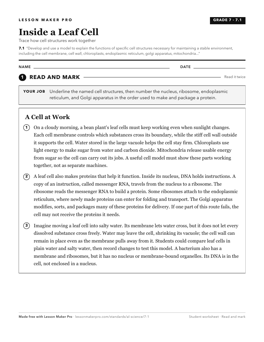
A ready-to-print activity worksheet aligned to 7.1, with an answer key for the teacher on its own page. No account needed.
PDF, US Letter, prints cleanly in black and white.
Download the worksheetKeep exploring
Related Standards
- BIO.3
Develop and use models to explain how events during the cell cycle lead to the formation of new cells and repair of multicellular organisms, including cell grow...
- BIO.4.a
Use models to illustrate how the structural characteristics of lipids and proteins in the cell membrane regulate cellular processes.
- HAP.1
Obtain, evaluate, and communicate information to explain how differences in cellular structure (mitochondria, cytoskeletal structure, endoplasmic reticulum, cel...
Turn this cluster into a lesson
Grade, subject, topic, and the complete cluster are prefilled. Create one free, no account needed.
Also for this cluster:Make a WorksheetMake a QuizMake a Sub Lesson